Cats
From Comparative Physiology of Vision
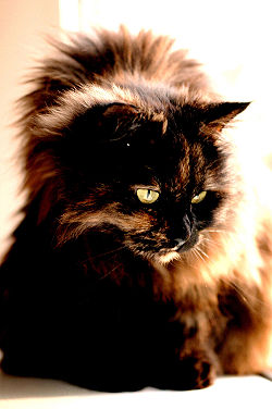
| Domestic cat | |
|---|---|
 |
|
| Longhaired Tortoiseshell Cat | |
| Scientific classification | |
| Kingdom: | Animalia |
| Phylum: | Chordata |
| Class: | Mammalia |
| Order: | Carnivora |
| Family: | Felidae |
| Genus: | Felis |
| Species: | F. catus |
| Binomial name | |
| Felis catus Linnaeus, 1758 |
|
| Synonyms | |
|
Felis catus domestica |
|
Contents |
Anatomical Schematic of a Cat's Eye
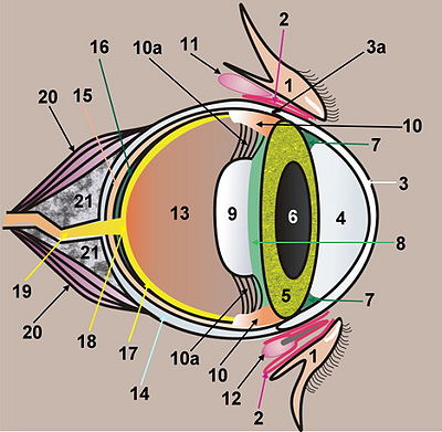
1. Eye lids Protects the eye from dust and abrasion.
2. Conjunctiva/Nictitating Membrane The conjunctiva is a mucous membrane covering the inner surface of the eye lids, and is connected to the sclera. The nictitating membrane may cover the entire front of the eye ball in cats.
3. Cornea The cornea is composed of different layers: the outer layer, the Epithelium, the Stroma, the Descemet's membrane, and the Endothelium on the inner side of the cornea. The cornea is transparent. It plays an important part in the refraction of the light rays. It has very fine nerve fibers spread out from the optic nerve which are responsible for the corneal reflex- involuntary closing of the eye lids in connection with an increased production of lacrymal fluid.
3a. Limbus Joins the cornea to the sclera.
4. Anterior chamber Located between the cornea and the iris, filled with the aqueous humour. The chamber is connected with the posterior chamber (8) through the lens (6).
5. Iris The eye's diaphragm, coloured by pigments. Located at the joint between the cornea (3) and the sclera (14). At its root it is connected to the ciliary body (10) and has the pupil (6) in its center.
6. Pupil The pupil is the opening within the Iris. It can be dilated or narrowed, regulating the intensity of the light rays reaching the retina. The adjustment of the pupil to different intensities of the light is called adaptation. Cats have a slit-like pupil, able to dilate or narrow the pupils much faster.
7. Canthus with Trabecular Meshwork The trabecular meshwork is a loose sponge-like tissue responsible for the drainage of the aqueous humour.
8. Posterior chamber Located between the iris and the lens, filled with the aqueous humor. This chamber is connected with the anterior chamber (4) through the pupil (6).
9. Lens The lens focuses the light to a sharp image on the retina (17). It does not have nerves or blood vessels- metabolism works through the aqueous humor (10). The lens is adapted to different distances by the suspensory ligaments (zonules) (10a) of the ciliary body (10), which means the lens can have different curvatures. If the muscle fibers of the ciliary body are relaxed, the ligament is taut, and the curvature of the lens is flattened so far away objects can be seen. If the muscle fibers of the ciliary body are contracted, the ligament is loosened, and the curvature of the lens is bent so closer objects can be seen. Adapting the lens to different distances is called accommodation. In humans the refraction index of the light rays is the same on the entire lens, but the lens of the cat is faceted into zones with different indices of refraction. This allows the cat to get a sharp image when the pupils (6) are narrowed to a slit.
10. Ciliary Body Connects the choroid (15) with the back side of the iris (5) and produces the aqueous humor which fills the anterior and posterior chambers (4, 8). The ciliary body has a suspensory ligament (zonules) (10a) connected to the lens (9).
10a . Suspensory Ligaments (Zonules) Responsible for the mounting of the lens and directing the curvature of the lens.
11. Lacrymal/Tear gland The lacrymal gland produces the lacrymal fluid. On top of the lacrymal film is a lipid layer produced by the Meibomian glands (1). The lacrymal fluid is drained off through the canaliculi into the lacrymal sac, and from there through the lacrymal duct into the nose. The lacrymal fluid has several important functions- protection of the cornea by draining debris from the eyes, lubricating the eye lids, protection against infections via antibacterial enzymes, removal of dead epithelial cells from the cornea, aiding in the metabolism of the cornea, and aiding in the transport of oxygen in the cornea. The lacrymal fluid has several layers: 1 On its surface is a lipid layer produced by the Meibomian glands (1) and the Zeis' glands. 2 The medium watery layer contains electrolytes and proteins produced by the lacrymal gland. 3 The inner mucus layer is produced by the accessory glands on the edge of the eye lids.
12. Nictitating gland The nictitating gland is an additional tear gland that produces about one third of the lacrymal fluid.
13. Vitreous Humor The vitreus humor is located between the retina (17) and the lens (9) to maintain the shape of the eye. It is connected through membranes. It consists of water and collagen fibers.
14. Sclera It builds the outer layer of the eye ball together with the cornea (3). Where the sclera joins the cornea is called the Limbus (3a), which is covered by the conjunctiva.
15. Choroidea Contains a network of blood vessels which are responsible for the retina's metabolism. It has several layers- the Lamina suprachoroidea (elastic ligament with pigmented ligament cells), the Lamina vasculosa (blood vessels), the Tapetum lucidum (16), the Lamina choriocapillaris (capillary network responsible for the metabolism of the retina), and the Lamina basalis on the pigmented layer of the retina.
16. Tapetum lucidum A reflective layer of the Choroidea (15) located behind the retina (17). The light rays are reflected back to the retina, amplifying the light signal. The part of the Tapetum lucidum which is non-reflective (the outer part of the tapetum) is called the Tapetum negrum.
17. Retina The image received by the eyes is analyzed and processed by the retina before it is transmitted to the brain. The light must pass through the layers of the retina before being received by the photoreceptors.
18. Optic Nerve Papilla The location on the retina of the optic nerve.
19. Optic Nerve
20. Eye Movement Muscles
21. Lipid Tissue
Visual Field
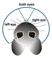
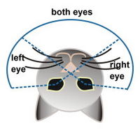
Being members of the family Carnivora, cats have adapted vision characteristics that enable to them to hunt prey. Cats have binocular vision similar to humans that allows them to focus on and catch prey. Prey mammals tend to have more developed peripheral and motion sensing vision than predators do, which allows them to quickly detect and react to stimuli rather than focus intently on one thing. The visual field of of predator and prey mammals also varies- prey tend to have a wide visual field with the eyes located on either side of the head, while predators have a narrower field of view that allows them the benefits of binocular vision such as increased depth perception and tracking ability.
Cats have a total visual field of 200 degrees, slightly greater than that of humans (180 degrees). Their binocular range, the area of the visual field with overlapping information from each eye, is 140 degrees, similar to that of humans. Despite this, cats are not as able as humans at focusing on near-field objects- their range of accommodation is limited to about 25 cm. If an object is closer than that distance, the cat cannot accurately focus on it. This explains why cats prefer to interact with close-range objects by touch or smell. Their highly sensitive whiskers aid in this close-range perception.
Color Perception
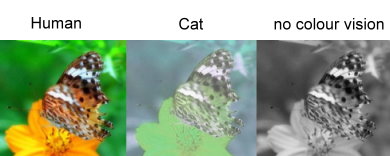
The ability to distinguish color is less pronounced in many predator mammals than prey mammals since color is not as important in the procurement of their sustenance as compared to detection of motion and boundaries. Prey animals, by contrast, often use color to detect the ripeness and identity of fruits and other food sources. It is theorized that this requirement is what ultimately led to the development of trichromacy in primates. Despite this, cats do have two types of color receptive cones that respond optimally in the yellow/green range and the blue range, making them dichromats. However, 83% of a cat’s cones are those in the yellow/green optimal receptive range, so their ability to distinguish and contrast colors is more limited than an animal with a more even distribution of cone types. Before the existence of the second cone was established, it was theorized that cats may have used their rod receptors, which respond optimally at a different frequency, to perceive color in conjunction with their cone receptors. The lack of a cone optimally receptive in the low-frequency visual range means that cats have a much greater ability to discern color gradients in the higher-frequency end of the spectrum (blue, purple, yellow, green) than in the low-frequency end of the spectrum (orange, red).
Visual Acuity and Sensitivity to LIght and Motion
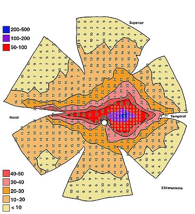
Cats lack a fovea and instead have a visual streak in the center of their visual field, which is known as the area centralis. The area centralis is the region of greatest photoreceptor (rod and/or cone) and retinal transductor (ganglion cell) density. The proliferation of photoreceptors in this region is an evolutionary adaptation for focusing and tracking. In humans, this high-density region of the area centralis is called the fovea. It is round in shape and contains nearly 100% cones, the high-resolution photoreceptors that allow us to perceive color. The visual streak in cats and other carnivores is similar in function but is slit-shaped rather than round. The two areas also differ in that the visual streak in cats contains primarily rod photoreceptors rather than cone photoreceptors. The density of rods in the visual streak of cats is about 463,000 per sqaure millimeter, and the density of cones is about 27,000 per square millimeter. Outside the area centralis, the density of photoreceptors is about 250,000 per square millimeter for rods and about 4,000 per square millimeter for cones. Humans, by comparison, have about 150,000 cones per square millimeter in the fovea and a rod density of about 150,000 per square millimeter at their maximum, which is at the periphery of the fovea.
The lower density of cones in the periphery of both humans and cats means that they must move their eye or head to maintain their focus on an object with any amount of visual acuity. It is suggested that the comparatively high density of rod photoreceptors allows cats to distinguish between finer shades of grey than species with lower photoreceptor density, but this is only true when the rods are not “saturated”. Rod saturation occurs when a great majority of rods have been activated due to high intensities of light. Once activated, rods must be “reset”, which takes time, before being able to perceive light again. This is what makes rods less effective than cones in high light-intensity conditions.
The low density of cones in the visual streak means that cats do not see as clearly as humans. The visual acuity of cats varies between 20/100 and 20/200 in the Snellen method of measurement, whereas most humans have 20/20 vision. This means that what a human could see clearly at 100-200 feet, a cat could only see clearly at 20 feet or less. This lack of acuity is partly compensated for by the proliferance of rod-type photoreceptors, which are more useful in night vision than cones due to their higher sensitivity. The high sensitivity of rods also confers greater ability to detect motion, a useful adaptation in tracking fast moving prey such as rodents. This makes cats very sensitive to lateral movement but not as sensitive to movement towards them, in the center of their visual field. This sensitivity to motion means that cats have a higher “flicker fusion rate” than humans, a measurement of how many identical images per second can be shown to an animal and be perceived as separate still images rather than one continuous image. The flicker fusion rate of humans is about 50 hertz (images per second), cats have a rate of about 70 hertz, similar to that of dogs.
Nocturnal Nature
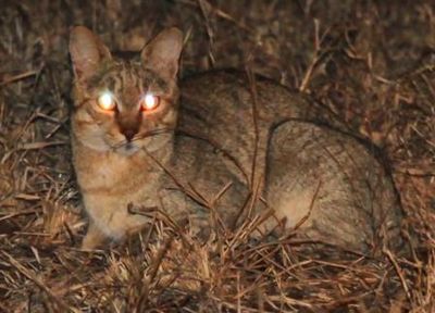
The high levels of rods in the retina of the cat is an indicator of their nocturnal status, meaning that they are adapted to hunt and catch prey at night. The high sensitivity of rods compared to cones is better suited to night vision. The nocturnal capabilities of the photoreceptors is further enhanced by the Tapetum lucidum, a thin layer of cells that the photoreceptors are embedded in. In humans, this layer is called the Pigment epithelium, and contains dark pigments to prevent the reflection of light within the eye. The Tapetum lucidum contains semi-transparent layers that reflect differing frequencies of light, which preferentially amplify some wavelengths of light as the refelections recombine. The wavelengths that result from this interaction depend on the original angle of the light as it strikes the tapetum lucidum and the compositional properties of the tapetum lucidum. This phenomenon is called iridescence, and explains why the eyes of cats appear reflective in low light conditions and why the appearance of this reflection changes if the cat moves its head in relation to the light entering its eye. In cats, the compositional properties of the tapetum lucidum are such that it tends to shift the light striking it to reflect light that is closer to the optimal receptive range of the rod photoreceptors in its eye. Although helpful in night vision, this iridescent layer can also have a disadvantage in that it decreases visual acuity during the day, which is why diurnal animals have a pigmented light-absorbing pigment epithelium instead. To compensate for this, the tapetum lucidum does not exist in the portion of the retina where objects in the upper periphery of the vision would strike. Instead, a darkly pigmented tapetum nigrum absorbs light in this region This is thought to prevent a bright, sunlit sky from iridescing in the eye and overwhelming the cat’s photoreceptors.
The lens of a cats eye is positioned closer to the optic nerve, which projects a smaller but brighter image onto the retina. The reduced size of the image results in decreased resolution but the brightness amplifies the signal, and important consideration in low-light conditions. Nocturnal vision in cats is also enhanced by their wide range of pupil dilation and constriction, allowing them to increase the pupil size to encompass nearly the entire cornea or decrease it to facilitate hunting during the daytime. In addition to this property, the cornea itself is of a large size compared to other animals which also increases the amount of light that can enter. The pupil size is also linked to the flight or fight response. The pupils of a cat dilate when it is threatened and becomes fearful to increase the light entering the eye, which allows for better detection of movement and facilitates the ability of the animal to escape the threat. If the cat instead responds aggressively to a threat, its pupils constrict to signal its intent to the offending animal.
Third Eyelid
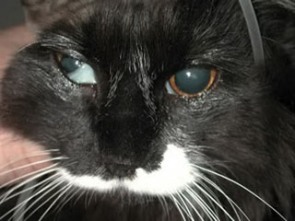
Although cats can “squint” to close their eyelids for protective purposes, they do not need to blink frequently to moisturize the eye. This allows the cat to focus on prey for a long period of time without breaking its focus or exhibiting motion which would give it away. Instead of moisturizing the eye frequently by blinking, cats use a “third eyelid”, known as the nictitating membrane, which covers the eye and moisturizes it. It also acts as a further protective layer, shielding the eye from damage. The nictitating membrane projects from the lower corner of the eye, moving across it diagonally to cover the eye as needed.
References
1. "Cat Eyes & Vision : Cat Guide : Animal Planet." Animal Planet. Web. 06 Dec. 2011. <http://animal.discovery.com/cat-guide/cat-anatomy/eyes-vision.html>.
2. Daw, N.W., and A.L. Pearlman. "Cat Colour Vision: One Cone Process or Several?" Journal of Physiology 201 (1969): 745-64. Print.
3. "Eye of the Cat." Katzenzeitung. Mar. 2007. Web. 06 Dec. 2011. <http://www.katzenzeitung.eu/en/Anatomy/Eye/index.html>.
4. Miller, Paul E. "Vision in Animals - What Do Dogs and Cats See?, by Dr. P. Miller - Alaskan Malamute Health." Alaskan Malamute Health Home Page. Web. 06 Dec. 2011. <http://www.malamutehealth.org/articles/eye_vision.htm>.
5. Schneider, Beller H. "The Spectral Sensitivity of Dark- and Light-adapted Cat Retinal Ganglion Cells." Journal of Neuroscience 13.4 (1993): 1543-550. Print.
