Fish
From Comparative Physiology of Vision
| Red Bellied Pacu | |
|---|---|
 |
|
| Scientific classification | |
| Kingdom: | Animalia |
| Phylum: | Chordata |
| Class: | Actinopterygii |
| Order: | Characiformes |
| Family: | Characidae |
| Subfamily: | Serrasalminae |
| Genus: | Colossoma |
| Species: | C. bidens |
| Binomial name | |
| Colossoma bidens (Spix & Agassiz, 1829) |
|
The Underwater World
General Setup of a Fish
Fish are vertebrates that must spend almost their entire life under water. This leads to some interesting differences in their anatomy than animals that live on land. They pack a lot of important abilities into what may seem to us like a rather "simple" looking body with fewer or less pronounced appendages than humans have. When eating them for food, it sure seems like they are one big piece of muscle, with a pair of eyes and lots of scales attached. But they are sophisticated!
Their ability to see underwater is interesting and will be our focus; they must deal with problems underwater that are not as severe in air. The refractive index of water is 1.33 for pure water, or possibly slightly greater for the kinds and temperatures of water fish can live in. Refractive index is a measure of the speed at which light travels through a material compared to a vacuum. This is a challenge because many species of fish must also be able to detect what is going on above the surface of the water, where the refractive index is lower. Whenever light moves from one medium to another, it bends and travels a slightly different direction if the refractive index is not the same. Another significant challenge to living underwater is Turbidity. Turbidity describes particulates in water can cause light to go back in directions it came from when it hits them rather than forward. More particulates equates to higher turbidity, and less light reaching the bottom of a body of water. Less light reaches the bottom because as the distance light travels from the surface increases, it has had more opportunity to run into suspended particles in the water and bounce.[1] Turbidity has much more affect on the quality and quantity of available light than refractive index.
The fish's eye is often found between the gill plate and the mouth, behind the nostrils. The location of the eye on a fish and the type of food it eats seem connected. Bottom dwelling fish who eat off the bottom or sift through sand may have eyes on the top of their head. Fish species that live in the middle of the water more commonly have the eyes on the sides.
Other major differences a fish has from mammals:
- Fishes do not have ears that stick out like mammals do.
- They can feel and hear vibrations through what is known as the "lateral line" on their sides.
- There are also ear-like structures in the head of the fish, but they are not open to the water. [2]
- Fish are very sensitive to underwater vibrations, this is why many aquariums have signs that say "Do not tap on the glass."
- Fishes are able to smell molecules that are floating around in water instead of air
- Humans have no hope of putting their head underwater and taking a good whiff.
- The nostrils possessed by bony fish are not used for for breathing.[3]
- Fishes must either conform to the environment outside of them, or regulate their insides carefully.
- They must manage to keep the proteins and ions in their body from drawing too much fresh water in.
- Taking on too much water without getting rid of it will make fish swell up.
- If a given type of fish lives in salt water, they probably have the opposite problem; they must keep the solutes in the ocean from drawing too much water out of their body.
- They must manage to keep the proteins and ions in their body from drawing too much fresh water in.
Please see http://en.wikipedia.org/wiki/Osmoregulation if you have an interest in this.
- Fishes possess gills that they use both for respiration and excretion; they eliminate nitrogenous waste from their gills in the form of ammonia.
General Anatomy
Fish have more spherical lenses than humans and, with few exceptions (sharks, rays), focus light on the center of their visual field by lateral movement of the lens—by moving it either toward or away from the retina, rather than by stretching the lens. The evolution of terrestrial vision has been linked to a common fish ancestor. Not surprisingly, the basic anatomical structures of the eye are similar to that of humans. Many fish possess rods and cones embedded in the retina that receives light as it passes through the cornea and the lens as well as a cone-dense fovea. However, many fish possess cone cells that are capable of absorbing wavelengths (WL) of light on the ultraviolet (UV) end of the spectrum. This feature, in addition to having two longer-WL receptors and one shorter WL receptor, makes some fish tetra-chromatic.
Despite the tetrachromacy of some fish, the kinds of light each species can absorb is highly variable. As a rule, a fish’s ability to see a broad range of color decreases with increase in the depth at which a given species resides. Trichromats, such as Pacific salmon, are generally surface dwellers whereas deep-ocean dwellers, such as the Flashlight fish, express photophores (cells capable of producing luminescence) in the retina which they use to effectively spotlight their prey. This unique trait enables flashlight fish to reflect a beam of light off of the tapetum (the tapetum is a feature shared with many terrestrial creatures, such as cats, which aids in night vision) of prey fish, which helps them assess the location of their fare. As no light reaches the deepest parts of the ocean, these fish do not see in color and rely on bioluminscence (provided by photophores) and rod cells to see.
Water Depth Between the broad range of colors available to the eyes of surface-dwellers and the complete lack of color vision available to deep sea fish, there exist many other levels of perceptive abilities available to fish at various depths. Anchovies can detect polarized light, which is most abundant at dawn and dusk. Since the sun travels from east to west, it is believed that this is an important trait in migratory fish as it may imbue a sense of direction. Polarized light-detection may also provide a sense of position in space relative to other fish in a school as polarized light is reflected off of the scales of neighboring fish. Reflection can also provide contrast with a diffuse background (a background that possesses no reflective properties). Contrast is an important property in fish vision and the implications range from relative location in space to camouflage (predator evasion), however this topic is beyond the scope of this article.
UV Light The ability of some fish to perceive UV light can serve as a means of prey detection. Juvenile brown trout absorb UV light as it reflects off of zooplankton, which is the staple of their diet. When brown trout mature and move into deeper water, they lose the cones that absorb UV light as this WL does not penetrate into the depths. This change in cone cell distribution is not unique to brown trout. Juvenile salmon also undergo a similar change as they move from the freshwater where they are born into the ocean where they mature.
Deep Water Deeper dwelling fish (1000M) typically have tubular eyes that face up and possess only rod cells, as very little light reaches these depths. Like humans, they have binocular vision. One of these types of fish, the barreleye, has a tough, flexible transparent dome covering their eyes, which is believed to protect the eyes from the “stinging” cells of their prey: the Man-of-War jellyfish.
Another type of barreleye, the brownnose spookfish, is the only species known to employ the use of a mirror-like surface to reflect an image on the retina. In essence, this fish, like the four-eyed fish, has the ability to look in two directions at once. The upward-looking eye is similar in function to other barreleyes, however its downward-looking eye utilizing a crystalline-structured mirror (possibly made of guanine crystals) to reflect light from below. This may seem surprising. However, recall that there is some form light in the depths: recall bioluminescence. The downward-facing eyes enable the spookfish to detect the bioluminescence produced by the photophores of the deeper-dwelling flashlight fish and other similar fishes.
Bioluminescence Yet another form of bioluminescence exists in the depths. The loosejaw fish possesses photophores that emanate infrared (IR) light. Its prey cannot detect this WL, so the loosejaw effectively searches for prey with an invisible flashlight. It seems that “uniqueness” becomes somewhat of an tired reference when referring to fish vision—particularly at great depths. Yet, back near the surface, swordfish demonstrate a striking trait. They have the ability to warm the temperature of their eyes (and their brain) up to 15 degrees Celsius. This increases the responsiveness of their eyes which aids them in keeping track of high-speed prey.
Eye Components and Photo Transduction
Eye Components
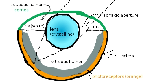
The above picture represents the eye of a fish. Light first passes through the cornea to enter the eye. The cornea is made up of layers of cells that make a tough, transparent skin.[4] When light leaves the water that fish swim in and enters the outside of the cornea it is bent slightly.
The cornea may contain iridescent cells that are aligned in such a way that light coming from certain directions is reflected back out of the cornea and away from the fish. This can dampen the effects of sunlight from above; glare is reduced by preventing light from specific directions from passing all the way through the cornea.
The difference in refractive index of water and cells of the cornea is similar on the inside and the outside. The initial bending of light caused by the cornea is undone as light enters the aqueous humor. The fish's cornea does almost nothing to contribute to refractive power of the eye because of this.
Light originating from an angle, like in this picture, utilizes the entire crystalline lens of the fish eye and is focused on a high resolution part of the retina. In species that are unable to change pupil size, there is an area in the eye known as the aphakic aperture. This is an empty hole that allows light to enter and hit the photoreceptors without passing through the lens. It enlarges the field of vision at the cost of image contrast. The cause of this is that light that has reached the retina through the aphakic aperture is not focused on the retina.[4]
In fish with irises that are able to change shape, the majority of light tends to make it through the lens, is focused, and hits the highest resolution part of the retina.
The retina is made up of neurons (pictured in gray), photoreceptors, and the sclera. Light travels through these components in this order. Because neurons and nerves are in front of the photoreceptors, light is lost. However, once light travels through the photoreceptors many species have a tapetum that reflects it back through the photoreceptors a second time.
Transduction (Work in progress)
Information from the photoreceptive cells must be passed to ganglion cells whose myleinated axons become the optic nerve.
These neuron cells do a lot of pre-processing:
There are horizontal cells directly in front of photoreceptor cells in the fish eye. These horizontal cells span a large area of the retina and release GABA neurotransmitter to cones and rods to inhibit their action in response to glutamate released from them. There are 4 layers of horizontal cells in most fish, and they are referred to as H1-H3 and "deep" horizontal cells. The job of the horizontal cells is to allow fish to extract "spacial contrast, rather than [respond] to the absolute level of illumination." [4].
In addition to horizontal cells there a few more important cell types, bipolar neurons, amacrine cells, and ganglion cells.
The bipolar neuron cells can be either transient (fire rapidly at first, then slow) or sustained (fire at constant rate that corresponds with stimulus intensity). They can accept input from both rods and cones, or one or the other. Depending on species, some of a fish's bipolar cells may hyperpolarize in the presence of one wavelength, and depolarize in another wavelength.[4] This is known as "double opponent" processing.[5]
The amacrine cells sit between the bipolar cells and ganglion cells. Amacrine cells can also be transient or sustained typed. The transient amacrine cells have huge dendrites that go literally everywhere in the retina, and the sustained amacrine cells have smaller more localized dendrites. The amacrine cells can act on the bipolar neurons to control their behavior.
Finally, ganglion cells extend dendrites towards bipolar and amacrine cells. It is the ganglion cells' axons that make up the optic nerve, and it is the Ganglion cell's on-center, off-center, or on-off fields that identify their receptive fields.
Unique Visual Optics
Eyelids
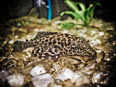
Fish do not have eyelids; their eyes are always open. Some fish may appear to "blink" when they roll/turn their eyes at a sharp angle. Interestingly enough the Plecostomus is a good example again. see http://www.youtube.com/watch?v=XOGNZwW2HF4&t=0m15s
Fixed Pupil
The pupil cannot constrict or dilate in many species.[6] This is probably because the turbidity of water in nature makes light an important commodity for many fish. It is a rather dark environment underwater, and fish have small eyes for their body size. [4]
Omega Iris
Not all fish are unable to adjust their pupil. For some fish, the shape and size of their pupil is dynamic and based on the amount of available light. One such fish is the Plecostomus, an algae eater that is available at any nearly any chain pet store. They possess a special kind of iris that at times looks a lot like the greek capital Omega symbol Ω. On the right side of this page you will find a picture as well as a rendition of the eye of a member of the Loricaria genus, which Plecostomus belong to. During night time, or in dark areas the iris contracts and more light enters the pupil. In the daytime or underneath the bright lights of an aquarium the iris of the plecostomus may expand until the iris looks more like the omega symbol.[7]
Polarized Light Detection
Many fish have cones that are somewhat connected to other nearby cones. One could say they're "joined at the hip."[8] According to the Encyclopedia of Fish Physiology, this pairing sometimes results in cells being "sensitive to the polarization of light by polarization-selective reflection and transmission of light" due to "cell membranes between the partners" making them this way.[4] This is not the only way fish can detect polarized light.
Another Means of Polarized Light Detection
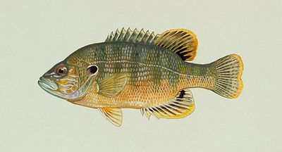
Some species have unique retinal cells that allow them to distinguish polarized light from the scattered background of the nonpolarized. In fish such as the green sunfish, a freshwater fish, the polarized light is detected through light vibrations as the waves of light move parallel to the axes of the cells in the eye. The sunfish have adjacent retinal cells that react to the polarized light with their abnormally large axes on the cell. The cells are all elliptical and perpendicularly arranged.[9]
Tapetum
Species that live at deep depths may have a tissue known as the "tapetum". The tapetum is behind the photoreceptors and is sort of like a mirror. It reflects certain wavelengths of light back through the retina so that the cells in the retina have an additional chance to get stimulated by the same light that entered. [4] This type of mechanism is found in nocturnal mammals and birds as well. [10] The effect created is known as eye shine. This eye shine effect is not the same as the red eye caused by camera flashes. Red eye is caused by light reflected by the optic disc and macula in the back of the eye. It's red because of the blood vessels back there. [11]
Example of eye shine in red belly pacu (take notice of the pink color!) http://www.youtube.com/watch?v=cvLEezA_Mz4 Same fish, different day: http://www.youtube.com/watch?v=hgr0o5O6TYY
Color Vision
Fish possess both rods and cones. The amount of photoreceptors and their type of visual pigments vary between species of fish. The environment the fish has evolved has a correlation with, but is not necessarily the cause of visual pigment types possessed by that species. Fish that live in deep water where little light reaches them may only have one or no cone types. There are four types of cone pigment opsins (a G protein-coupled receptor) that fish may have. A fish with all four types is known as a tetrachromat. These four cone types all first evolved in vertebrates almost ~450-500 million years ago.[4] Two types of cone opsins available to fish are not found in mammals. Humans have two versions of the LWS cones (which allow for humans to distinguish "red" from "green".[12]
Cone Types:
| RH2 MWS cones | SWS2 SWS cones | SWS1 UVS/VS cones | LWS LWS cones |
| 470-530 nm wavelength | 415-480 nm wavelength | 355-450 nm wavelength | 495-570 nm wavelength |
| fish, amphibians, birds | fish, birds | fish, amphibians, mammals, birds | fish, amphibians, mammals, birds |
It is not the intensity of response among a single cone type in the retina that allows fish to see in color, but rather the difference in responses of receptors that are more sensitive to different wavelengths of light that makes color vision possible.
Motion Detection
Detection of motion is an immensely important capability for any organism in the animal kingdom. Fishes utilize the ability to detect motion in their environment as they swim in order to avoid predators, catch prey, locate nearby fishes, and find mates and shelter. Fish are able to do this via their optomotor response - which allows them to maintain a gaze on a fixed object while their body is in motion - and the optokinetic response - which enables fish to detect movement in its surrounding environment.
Optokinetic Response
Researchers, in a study of zebrafish, have found that vertebrate motion processing is quite similar to that of primates. The pretectal area (APT) of the brain, which sits between the thalamus and midbrain, contains a region of neurons which receive binocular sensory input in fish. In mammals the pretectal area is involved in pupil reflex. [13] In teleost fish, direction-selective neurons in the APT are most important in global motion detection, most notably in controlling slow phase eye movement during gaze stabilization. A study carried out by Masseck, et. al. testing global motion detection thresholds in goldfish concluded with findings of very high levels of sensitivity for global motion in the optokinetic system. [14]
Optomotor Response
The Optomotor response has been found to require a much greater level of stimulation (sometimes twice to four times as much) in order to be triggered in fish. [14] The tectum opticum has been the most notable region of involvement in the optomotor response. Some researchers have presumed that the differing thresholds between the optokinetic and optomotor response indicate that the direction-selective neurons in the APT that are active in the optokinetic response are not involved in the optomotor response.
Neuronal Processing
Research on goldfish in boxes has shown that the fish pretectal area is responsible for motion detection and that there is a lack of control from the cortex. [14] Fish see and respond to movement very reflexively.
Higher order processing
Depth, Wavelengths, Lures and Baits
- On a bright day in clear water, the best lure to be using is the brightest one possible.
- I speculate that it is because there is plenty of light available to reflect off it and be seen by fish.
- On a cloudy day one should use a dark colored lure. An all-black lure would be great, because with less light comes less use of cone (color sensitive) photoreceptors, and more reliance on rods. For a fish, a dark shadow passing over it in the water will look like good prey.[15]
- Red light is lower in energy (frequency) than green and blue light. This makes red lures fade away into the shadows or look black in deeper water. To catch fish in deep water use a reflective lure or white one.
- It would be really good to use complimentary colors for baits to show something that has high contrast to catch a fishes attention. Like humans, fish are sensitive to "edges".
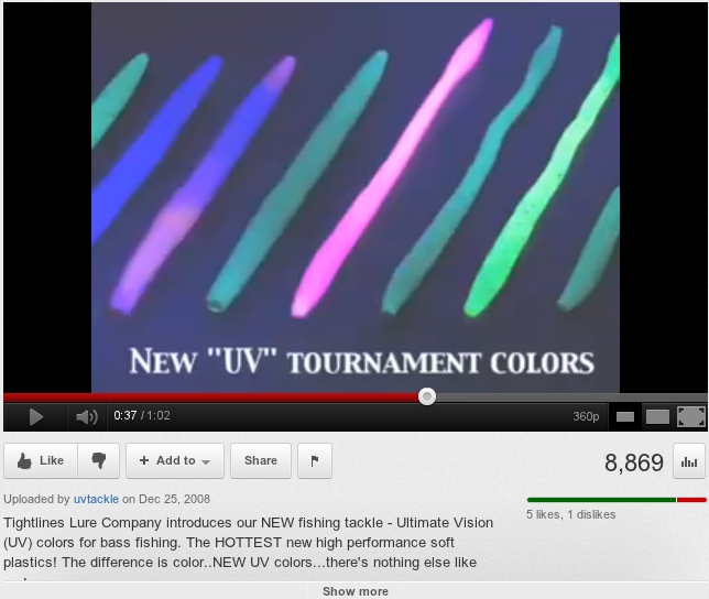
- If the species you are trying to catch is known to be interested in UV http://www.uv-tackle.com/tightlines_history.php
Luminous paint
Lures can be painted with two kinds of paints that give off light, they are good for deep water use, and nighttime use.
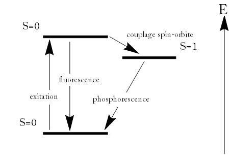
An older type of paint for both deep water use and nighttime use is phosphorescent. The electrons in the paint are excited and hang out in a higher energy orbital than normal for some time, before dropping back to old, lower energy level orbitals where they were before. As they drop back, a photon of light is released. This is a slow process and the bait gets dimmer and dimmer over time.[16]
There are also fluorescent lures, which absorb UV and give off a different wavelength of light.
A newer type of paint is incorporated into "UV tackle" (see http://www.uv-tackle.com/tightlines_history.php) and this kind of paint simply reflects UV light from as far as I can tell. http://www.youtube.com/watch?v=4_AoM-rLUD4 is video of it in action. To the fish, the UV Tackle is very bright compared to everything around.
Cool and Strange Stuff
Flatfish
- There are fish that hatch out of their egg with eyes in normal places like a normal fish, and over the days following their hatching one of the eyes "moves" to the other side of the fish. See this video: http://www.youtube.com/watch?v=qePwW44HhNg.
- The "blind" side of the fish is white and the lack of pigment helps hide the flatfish if it leaves the bottom and swims over other fish.
Four eyed fish
- The anableps has a double cornea, double iris(pupil), and retina with two distinct sections. The fish spends most of its time at the surface of slow moving water, looking for food.
- The top half of the eye is held out of the water and has a flat lens[17]
- The middle of the eye is set up with pigment to block glare from the surface.
- The bottom half of the eye is below the water and the lens is dramatically curved[17]
- The retina were at one time were thought to be all the same, and was mentioned this way in Scientific American on March 23rd, 1878.[18] It's now known there are two distinct sections of the retina in the anableps that feed into the optic nerve separately.
- The population of cones responding to light from above the water and below the water is different.
- The double eye is a result of gene duplication where both versions keep working, duplicated gene isn't a dud. [19]
Sharks
- Some sharks have an "eyelid" called a nictitating membrane. It's a translucent bit of skin that slides across the eye, offering the cornea some protection[20]
- Humans do not have nictitating membrane, like sharks. The vestigial (originated from but now useless) structure we have is a small flap made of epithelial cells (called "Plica semilunaris of conjunctiva" if you really want to know) at the inside corner of our eyes [21].
- The nictitating membrane is rare in fish
Tidbits
- As mentioned, some fish posses a tapetum, but can also disable their tapetum by forming a dark pigment layer over it when bright conditions are present.
- As there is litle difference between the cornea and water as far as refractive index is concerned, the lens in chiefly responsible for refraction[22]
- In contrast with most other fish, sharks, rays, and certain species of catfish do have a muscle attached to their iris, which enables them to adjust pupil size to focus light. See our section on omega iris.
- Some fish capable of absorbing UV light also reflect UV light off their bodies as a means of signaling other fish of predators that are not sensitive to UV light.
- Some fish possess double-cones connected by gap junction (enable free flow of ions and molecules between cells), however little is known of the purpose of this trait.
- There is at least one known “four-eyed” fish (Anableps). But this fish doesn’t wear glasses. In reality, it possesses two eyes divided into two separate compartments, each containing a pupil of its own. * One surface of each eye has adapted to the refractive index of air and remains above the surface of the water; the lower part of the eye remains submerged and is adapted to the refractive index of water. * Some of their prey is found on the surface, while some is found beneath.
Evolutionary Significance
Pupillary Light Reflex
The Pupillary light reflex is absent in some fish. It may be that being able to constrict the pupil did not give all fish a selective advantage, and thus there are fish without this feature. The reflex is unneeded in deep water or water with high turbidity.
Tapetum
The red belly pacu we have video of showed red colored light being reflected back through the retina. This kind of fish likes the company of one or more of its own species when it is a juvenile. As this type of fish ages, it loses its red coloring and no longer seems to care about having company as much. Could it be that the tapetum allows this fish to find others of its kind in the muddy waters of its natural habitat (Amazon River) or is it simply the color of sunlight that we see bouncing back out of the eyes of this fish?
Bioluminescence
Some fish evolved the ability to release electromagnetic energy to the environment, and detect (or lure) other fish that the energy is exposed to. This is most advantageous to fish who live where it is very dark.
Recent Discoveries
Importance:Findings led by a research team at Emory University, led by Shozo Yokoyama, has strengthened the importance of adaptive evolution. This realization regarding this species is highly significant because it is the first example of an animal actually deleting a molecule to change its visual spectrum; the deletion of this molecule occurs at the site 86, which is located on a chain of amino acids in the opsin molecule. It was discovered that the scabbardfish(Lepidopus fitchi) is currently the only known fish to have switched from ultraviolet vision to violet vision;the ability to see blue light. Another discovery scientists found in this species is that unlike previously studied fish, Scabbardfish do not have retained UV vision. Likely Cause: Yokoyama theorizes this shift occurred because they spend a great deal of their life at 25 to 100 meters, where UV light is less intense than violet light.
Notes and References
- ↑ http://en.wikipedia.org/wiki/Turbidity
- ↑ http://www.britannica.com/EBchecked/topic/208456/fish/63645/Hearing
- ↑ http://www.sbs.utexas.edu/shankland/lc15rsp1.htm
- ↑ 4.0 4.1 4.2 4.3 4.4 4.5 4.6 4.7 Encyclopedia of Fish Physiology: From Genome to Environment, Academic Press, 2011.
- ↑ http://en.wikipedia.org/wiki/Color_vision
- ↑ http://www.advancedaquarist.com/2010/3/fish
- ↑ http://en.wikipedia.org/wiki/Loricariidae
- ↑ http://www.vision-in-cichlids.com/cichlid_eye.htm
- ↑ http://discovermagazine.com/1996/oct/throughafisheyel905
- ↑ http://www.owls.org/Information/eyesight.htm
- ↑ http://en.wikipedia.org/wiki/Red-eye_effect
- ↑ http://www.diycalculator.com/sp-cvision.shtml#A7| Clive (Max) Maxfield and Alvin Brown
- ↑ http://en.wikipedia.org/wiki/Pretectal_area
- ↑ 14.0 14.1 14.2 http://www.plosone.org/article/info:doi%2F10.1371%2Fjournal.pone.0009461
- ↑ http://texastacklefactory.com/index.php?option=com_content&view=article&id=5&Itemid=111
- ↑ http://en.wikipedia.org/wiki/Fluorescence
- ↑ 17.0 17.1 http://www.internal.schools.net.au/edu/lesson_ideas/optics/optics_wksht4_p1.html
- ↑ http://books.google.com/books?id=WIo9AQAAIAAJ&pg=PA184&lpg=PA184&dq=double+iris+retina+fish+anableps&source=bl&ots=hoH1xbwIMs&sig=v3AzWdxc1IQM6_HXmevlZnRK-xY&hl=en&ei=nZncTvCpKobq0gHQkuXkDQ&sa=X&oi=book_result&ct=result&resnum=1&ved=0CBwQ6AEwAA#v=onepage&q=double%20iris%20retina%20fish%20anableps&f=false
- ↑ http://www.sciencedaily.com/releases/2011/07/110720142133.htm
- ↑ http://en.wikipedia.org/wiki/Nictitating_membrane
- ↑ http://en.wikipedia.org/wiki/Plica_semilunaris_of_conjunctiva
- ↑ (Land, M.F. ; Nilsson, D. (2002) Animal Eyes. Oxford University Press).
23. http://www.sciencedaily.com/releases/2009/10/091016121827.htm
Questions for class
1. (Short Answer) Describe how some fish (members of the Loricaria genus) change their iris to the shape of the Greek symbol Ω. When do they do this?
2. As measured ___________ increases, there is a significant loss of quality and clarity of light reaching fish underwater:
- a. refractive index
- b. pollution
- c. turbidity
- d. density
- e. lens
3. (Short answer) Briefly describe one way a fish might detect the polarization of light?
4. Question 4 (If you would like, replace one of our other questions) : What is the advantage to the ability of the brownnose spookfish to see down into an increasingly diffuse environment?
